
What is heart and how it works?
Heart is an important organ of our body. It is responsible for the flow of blood through circulatory system. The Circulatory system consists of
1. Heart
2. Blood vessels
3. Lungs
HEART
Heart is the great pumping organ, maintaining the circulation of blood through body.
BLOOD VESSELS
Blood vessels are of three types.
(i) Arteries
Arteries carry blood away from the heart.
(ii) Veins
Veins carry the blood back into the heart.
(iii) Capillaries
Capillaries connect the arteries & veins.
Lymphatic system also plays an important role in the circulation of blood. Lymphatic vessels collect the fluid (lymph), which was left from the circulatory system .
HEART
SHAPE
The heart is cone or pyramid shaped. It is a hollow muscular organ. The weight of heart is about 250 to 300 grams in male and 200 to 250 grams in female. The length of heart is about 12 cm , width is about 9 cm & thickness about 6 cm. It has four borders i.e upper, lower, left & right and are three surfaces, anterior, posterior, & diaphragmatic. The size of heart is about a closed fist.
POSITION
The heart lies in the thorax, within the pericardium, between the lungs & behind the sternum. A line drawn from the 3rd right costal cartilage, 12.5mm from the sternum, upwards to the second left costal cartilage, 18mm from the sternum, marks the position of the base of the heart. A point on the left side in the 5th intercostal space 9 cm from the mid line, gives the position of the apex of the heart. By uniting these markings by lines, as shown in the diagram, the position of the heart may be indicated.
STRUCTURE OF THE HEART
EXTERNAL STRUCTURE
The heart has four chambers, right & left atriams and right & left ventricles. Atria are above, while ventricles are below. On the surface of the heart, atria are separated from ventricles by intera-ventricular groove. The atria are separated from each other by inter-atrial groove, while ventricles are separated from each other by intra-ventricular groove. The heart is externally covered by visceral pericardium (epicardium).
(i) RIGHT ATRIUM
It forms the right border, part of the upper, interior and posterior surface. It receives the superior vena cava at the upper and inferior vena cava at lower end. The upper end is prolonged to the left to form the right auricle.
(ii) RIGHT VENTRICLE
It is triangular chamber. It receives the blood from the right atrium and pump it into the lungs through pulmonary trunk. Pulmonary trunk is funnel shaped called infundibulum. It forms the lower border, anterior surface and small part of diaphragmatic surface.
(iii) LEFT ATRIUM
It is quadrangular chamber. It forms the posterior surface, part of anterior surface & left border. It has a main cavity and a left auricle. It receives four pulmonary veins from the lungs.
(iv) LEFT VENTRICLE
It forms apex of the heart, anterior surface, left border and diaphragmatic surface. It pumps the oxygenated blood into the aorta for circulation.
INTERNAL STRUCTURE
The heart is internally covered by a layer of endo thelium (squamous epithelium), called endo cardium. The walls of the heart are composed of cardiac muscle called myocardium. The ventricles have the thickest walls. The walls of left ventricle are thicker than the right ventricle. The atria are separated from each other by atrial septum, while ventricles are also separated by ventricular septum.
(i) RIGHT ATRIUM
The main part of the right atrium is smooth walled called sinus venarum. The remaining part is rough containing, bundles of muscle fibers, called the "musculi pectinati". The superior & inferior vena cava empty their blood into the right atrium.
(ii) RIGHT VENTRICLE
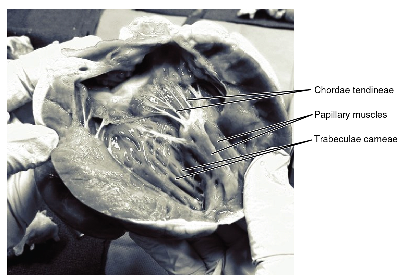
The right ventricle communicates with right atrium, through ventricular opening guarded by tricuspid valve, the inflowing part of the right ventricle is rough, due to muscular ridges, called "trabeculae carneae". The out flowing part is smooth called infundibulum.
(iii) LEFT ATRIUM
The interior of the left atrium is smooth, but the auricle has muscular ridges, called "musculi pectinati". Four pulmonary veins enter the left atrium, having no valves.
(iv) LEFT VENTRICLE
It communicate with left atrium through left atrio-ventricular opening having mitral valve (bicuspid) and with aorta through aortic opening having aortic valve (tricuspid). The Walls of the left ventricle are three times thicker than the right ventricle. The upper part is smooth gives origin to aorta called aortic vestlbule. The lower part is rough contains "trabeculae carneae".
FUNCTIONS OF THE HEART
- Heart maintains circulation of blood in the body.
- Contraction of heart maintains and regulates blood pressure.
- Heart keeps oxygenated & deoxygenated blood separate from each other.
- Heart contains valves, which prevents back flow of blood.
- Heart receives deoxygenated blood from Whole body & sends it to lungs for oxygenation.
- Heart Receives oxygenated blood from the lungs & distribute it in the body, for oxygen & nutrient supply to the body etc.
BLOOD SUPPLY OF THE HEART
(i) Arterial supply
(ii) Venous drange
NERVE SUPPLY OF THE HEART
The heart is innaired by sympathetic (cervical and upper thoracic) and para sympathetic fibers of the autonomic neotous cardiac plexus.
CIRCULATION OF THE BLOOD
Systemic (General) circulation
The blood is circulated from left ventricle, through the arteries, arterioles & capillaries. Then from the capillaries to venules, veins, returning into the right atrium, is called greater or systemic circulation.
Lesser or Pulmonary Circulation
The course of the blood starts from the right ventricle, through the pulmonary trunk, into the lungs and from the lungs, through the four pulmonary veins, into the left atrium, is called the lesser of pulmonary circulation.

0 Comments